Transforming pre-operative planning
Dramatically improve patient care with our proven patient-specific solutions
Surgeons typically rely on 2D patient scans to aid in diagnosis and pre-operative planning
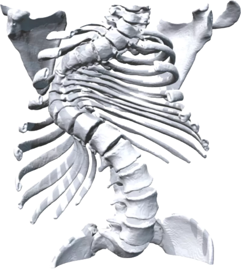
These commonly used 2D images make conceptualizing complex three-dimensional anatomical structures a difficult task for even the most experienced surgeon.
2D images can complicate pre-operative planning, leading to many complex surgeries being misdiagnosed or misplanned and millions of unnecessary hours spent in surgery.
These images can be hard for patients to decipher too, and surgeons will often have to resort to drawings and other tools to help the patient understand their diagnosis and to gain their consent to proceed with a planned surgery.
From planning to treatment – we make all cases patient specific
• Pre-operative planning
• Surgical simulation
• Intrateam discussions
• Gaining patient consent
• Reduced time and cost of surgery
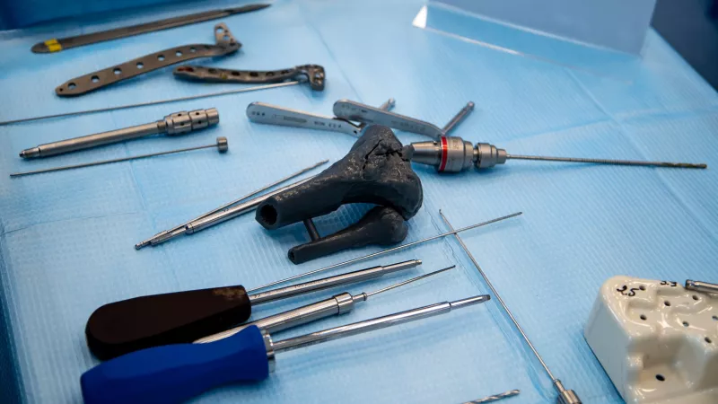
Patient-specific pre-operative planning is the future
References can be found here.
"During the course of work up of the donor, Computed Tomography imaging revealed a complex cyst (lesion) within the renal cortex. [...] As the cyst was buried deep within the renal cortex and therefore invisible on the back bench, a replica 3D model was used for pre-operative planning and intraoperative localization of the lesion. It’s difficult to underestimate how valuable this strategy was in terms of pre-operative planning and achieving successful clearance of the lesion."
Experience Axial3D for yourself
Have an existing or previous complex case? Request a patient-specific model of your patient's case to dramatically improve care at your facility.


 View a sample 3D model
