3D Anatomical Model Aids Surgical Planning in Severe Scoliosis Case
Scoliosis is a common spinal condition frequently diagnosed in adolescents. Each year, approximately 3 million scoliosis cases are reported in the United States, with a significant number being idiopathic scoliosis, which typically presents in children aged 10 to 12.
Case
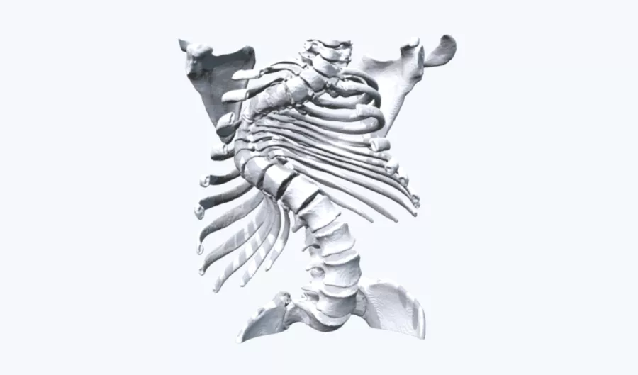
A 13-year-old male presented with congenital scoliosis, a condition characterized by an abnormal curvature of the spine present from birth. The complexity of his scoliosis made it challenging to accurately assess the curvature of his spine and understand the spatial relationships between his vertebral column and vital organs. Traditional imaging methods struggled to provide a comprehensive view of the intricate anatomical details required for precise surgical planning.
Solution
To address these challenges, Mr. Andrew O'Brien, Spinal Surgeon, utilized a 3D printed model from Axial3D, which was created from the patient's CT scan data. This model allowed for a thorough evaluation of the patient's condition, providing a clear and detailed representation of the spinal deformity and its impact on surrounding structures. Additionally, the model served multiple purposes: it was used to measure and prepare surgical equipment, ensuring accuracy and readiness before the operation, and it acted as a valuable educational tool for both the patient and his parents. This hands-on approach facilitated better understanding and communication of the surgical plan and its implications.
Benefits of the 3D Printed Model
With access to the 3D printed model, the spinal surgeons worked closely with thoracic surgeons to define a surgical plan. With the scapula and the ribs printed in 1:1 scale along with the spine, they could clearly understand the anatomical relationships. The model also allowed the planning of screw locations and trajectory into the pedicle of the spine, almost impossible to visualize and plan using CT images alone.
“In addition to allowing us to create a detailed surgical plan, the model also allowed us to convey the level of the complexity to the parents and set their expectations for anticipated surgical outcome.“
- Mr Andrew O’Brien, Spinal Surgeon
This 3D anatomical model was printed at 1:1 scale and delivered in less than 48 hours.
Disclaimer: Details of Axial3D's regulatory clearance for diagnostic use cases are outlined here. For all other uses of Axial3D solutions, they should be used for demonstration and education purposes only.
Discover how 3D printed models can help enhance your pre-surgical planning.
Talk to an expert.


