Enhancing Cardiac Imaging with 3D to Achieve Personalized Medicine at Scale
In today’s healthcare landscape, groundbreaking technologies like wearable devices, remote monitoring solutions, and the integration of AI and machine learning are driving patient-centric care like never before.
These innovations not only improve engagement and empower patients and their loved ones, but also improve clinical outcomes across the continuum of medicine. They now play a pivotal role in precise risk assessment, early disease detection, and the formulation of personalized treatment strategies.
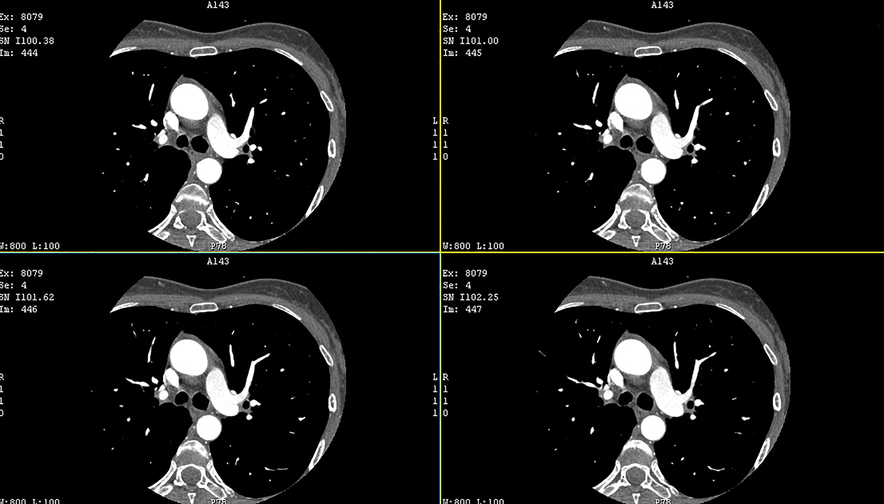
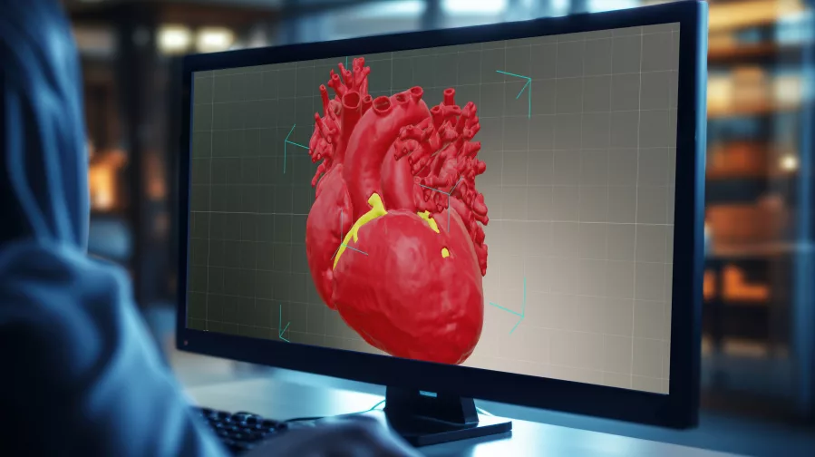
AI-driven machine learning algorithms, which are capable of analyzing extensive datasets quickly and accurately, are becoming an indispensable tool in clinical settings to improve device selection and fit by creating 3D data from DICOM images—particularly in cardiovascular medicine.
The Limitations of 2D Cardiac Imaging

Accurate cardiac imaging, which is crucial for the correct diagnosis and management of disease, can become a barrier to precision care for a number of reasons. First, the nature of the continual movement of the heart can create artifacts, making it difficult to obtain clear, interpretable images. Capturing the heart’s dynamic functions, including systolic and diastolic performance, valve movement, and blood flow, can be complex.
Various factors can also impact image quality and obscure valve and wall motion details including patient movement during the scan, obesity, lung disease, or previous surgeries.
Not only that, but the traditional methods of interpreting medical images are subject to variability and a margin of error. Distinguishing between normal anatomical variants and pathological findings requires significant expertise—and is time-consuming for both radiologists and cardiologists.
The New Frontier of AI-Driven 3D Imaging
AI algorithms can quickly and accurately analyze 2D images, identify abnormalities and provide detailed insights in 3D that can aid in diagnosis and treatment planning.
Analyzing DICOM images at scale. AI algorithms can analyze DICOM images quickly and accurately to determine the precise dimensions and characteristics of the cardiovascular structures. For patients requiring heart valve replacements, AI algorithms can analyze images of the heart to determine the exact dimensions of the valve annulus, ensuring that the prosthetic valve fits accurately and functions effectively.
Selecting and fitting medical devices. The benefits of AI and ML in cardiovascular care extend to the selection and fit of devices like stents, pacemakers, and heart valves. Ensuring that these devices are optimally sized is critical to minimize the risk of complications such as stent thrombosis or restenosis.

Predicting patient outcomes and stratifying risk. AI models can analyze patient data to predict the likelihood of adverse events, such as heart attacks or strokes, prompting preventive measures. These models can also identify patients who are most likely to benefit from specific treatments, enabling personalized care plans that improve outcomes and reduce costs.
Customizing medical devices. AI algorithms can be used to create patient-specific models of the heart, which can then be used to design custom-made devices at scale. This level of customization is particularly important for patients with complex anatomies or unique physiological conditions, as it ensures that the devices provide optimal performance and durability.
At Axial3D, our mission is to make patient-specific care routine. What does that look like in practice? Learn more about how healthcare organizations and medtech companies can leverage AI to build your patient-specific programs at scale.


