Case Study18th January 2024
Revolutionizing Pediatric Cardiac Care at Southampton Hospital
Improving pre-operative planning with anatomical 3D modeling: Removing a maxillary sinus tumor
Case Study
The Case
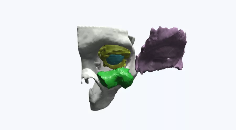
An 85-year-old male presented with a large lower-eyelid, cheek and nasal tumor. The tumor's size and location was causing both orbital invasion and maxillary invasion and needed surgical intervention. Working with 299 CT images from the hospital, Axial3D produced two 3D printed models and a 3D visualization to support pre-operative planning for the maxillectomy procedure.
The Solution
The first 3D printed model was used to pre-bend the plate of the orbital floor ahead of surgery, while the second 3D printed model was used to support intra-team communication between the cranio-facial and neurosurgery teams.
The Benefits of using a 3D Model
Having access to the precise 3D printed patient models significantly enhanced the planning process for the tumor surgery, fostering consensus and eliminating discrepancies among the cranio-facial and neurosurgery teams. The integration of the 3D models resulted in substantial time savings of approximately 80 minutes, enabling the development of a robust pre-operative strategy. Utilizing Axial3D's comprehensive models empowered clinicians to meticulously plan and execute procedures, ultimately improving patient care and ensuring comprehensive tumor removal.
Try Axial3D with a free personalized 3D anatomical model.
Request a free anatomical visual
Case Study8th November 2023
Enhancing Surgical Precision: Patient-Specific 3D Model for Vascular Calcification Assessment
Case Study28th August 2023
Optimizing surgical outcomes with 3D models in Double Outlet Right Ventricle (DORV) cases
Case Study28th August 2023
Gaining insights into anatomical intricacies of ccTGA using patient-specific 3D solutions
Case Study2nd December 2021
Correcting Transposition of the Great Arteries with the Help of a Patient-Specific 3D Printed Anatomical Model
Case Study29th November 2021
Medical 3D model saves crucial time in heart transplant surgery for a patient with congenital heart disease
Follow us


