
As a child, Dr. Furdova was diagnosed with septal defect following some check-ups on her heart. Small atrial septal defects can spontaneously close during childhood, however this is not always the case.
“During my studies I became very interested in cardiac surgery. Before taking on a new position, during a routine check-up, my GP referred me to a cardiologist due to a loud heart murmur. My cardiologist found a hemodynamically significant large atrial septal defect, and changes on my heart including some tricuspid regurgitation, right sided heart enlargement, and signs of pulmonary hypertension.”
At that moment, everything suddenly clicked for Dr. Furdova. She had previously experienced some difficulties with breathing and found it hard to stand for long hours, which of course caused great difficulty in her role as a cardiac surgeon resident, where a typical procedure can be anywhere between 4-6 hours long.
Dr. Furdova explained, “This all happened during my first year in cardiac surgery and it was so surreal. I was a student in this field, I am a doctor there now, and I was to become a patient!”
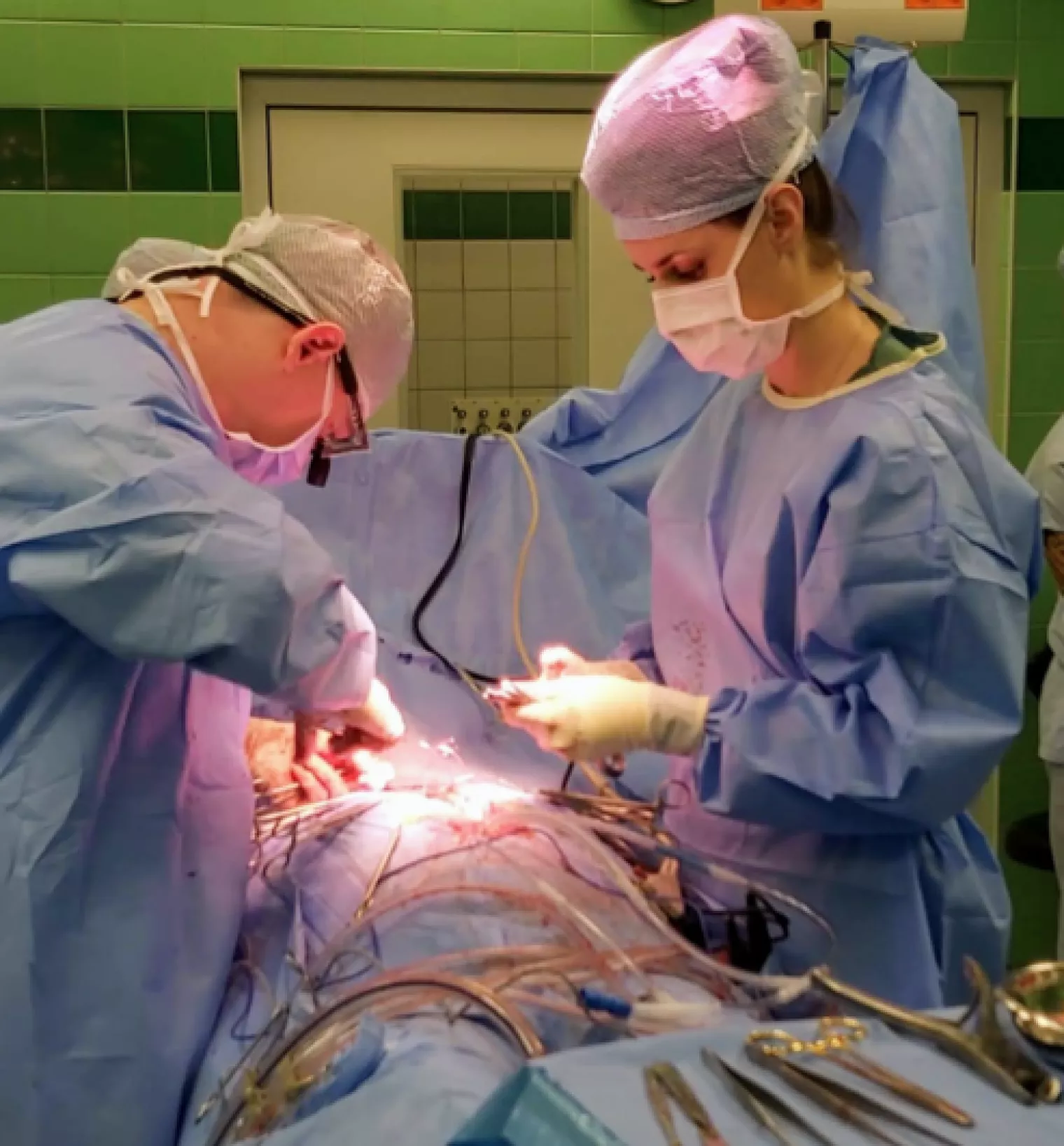
Being a cardiac surgeon, Dr. Furdova was very aware of the risks involved in the upcoming Da Vinci approach procedure.
“I became really scared knowing the possible complications. I had to come to terms that I would have to stay in the hospital for the first time in my life. I’d have to face the long operation for six or seven hours. I’d have to face the anesthesia. I thought to myself, ‘if I can survive this, I can survive anything’.”
Thankfully, Dr. Furdova’s procedure was successful and she’s now back at work treating patients who are in a similar position to the one she found herself in just last year.
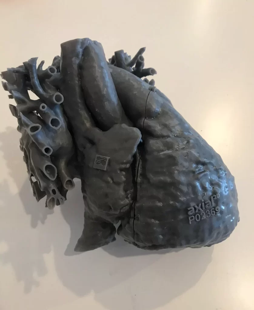
Discussing the day that her own 3D model with the atrial septal defect arrived on her ward, Dr Furdova says, “I was so enthusiastic, I must have shown it to everyone on my ward. No one could believe the size of the heart itself, and of the ASD, that it wasn’t a valve!”
“Seeing my own defects presented in such an accurate way on the 3D model was amazing. I think that patients would be really enthusiastic about having those models too, especially for special structural cases where they cannot understand it. It gives them as close as possible, an understanding of the surgeon’s perspective.”
The additional insight provided to surgeons by these precision 3D models can translate into a reduction in operating time and costs, explains Dr. Furdova.
“Sometimes surgeons are unable to understand all the details of a disease’s pathology even in 3D echocardiography. Having an accurate model helps you to perfectly prepare and measure everything ahead of the surgery. It helps you to understand the position of the cardiac structures in a real way and because it’s a physical model, you can touch and interact with it. The more prepared you are in terms of your surgical plan can mean a much shorter time in the operating room.”
“Axial3D is incredibly focused on providing surgeons with the details they need from the models. It doesn’t feel like it’s just ‘business’ for the team. You can tell there is a genuine care about the relationship between the surgeon and the patient.”
“And of course, time is precious. The segmentation time is so valuable because sometimes patients cannot wait for four months on a model. They need one within a few days which is what Axial3D delivers.”
Dr. Furdova is a Resident in a Cardiac Surgery Department in Prague. She has experience in the healthcare startup environment and has built a library of anatomical heart models called 3D Hearter. As a General Doctor she has experience of working in Africa (South Sudan) within a humanitarian program. She is passionate about improving healthcare globally and an advocate for social well-being.
