3D Anatomical Model Aids Surgical Planning in Severe Scoliosis Case
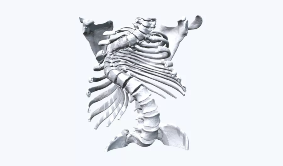
Scoliosis is a common spinal condition frequently diagnosed in adolescents. Each year, approximately 3 million scoliosis cases are reported in the United States, with a significant number being idiopathic scoliosis, which typically presents in children aged 10 to 12. Case A 13-year-old male presented with congenital scoliosis, a condition characterized by an abnormal curvature of the […]
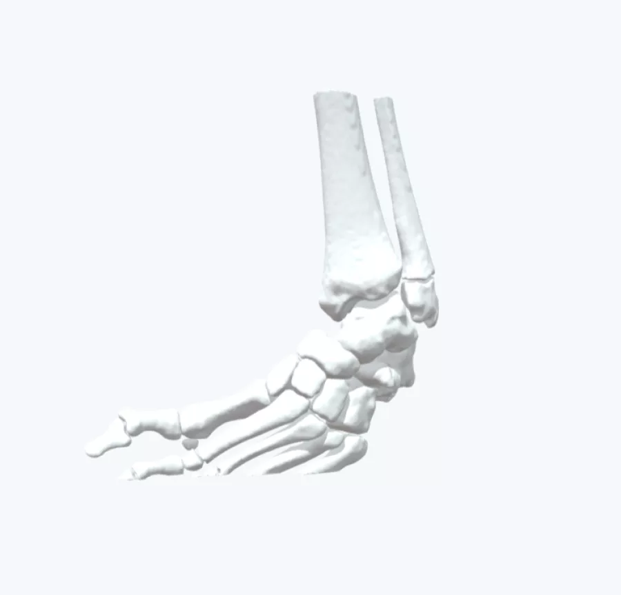
Utilizing 3D modeling to better understand a complex ankle trauma fracture

This 60-year-old male’s CT scans presented a complex ankle trauma fracture which required surgical intervention to enable his safe recovery back to full health. In planning, his surgeon was concerned that the CT scans alone did not provide him with a full picture of the problem, making it difficult to form an appropriate, definitive operative […]
3D printed twisted radius and ulna model reduces surgery time by 3.5 hours
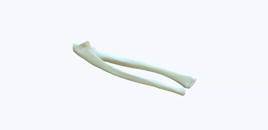
A patient-specific physical 3D printed model provided better insight into the pathology leading to a drastic reduction in theatre time and optimum result for the patient. Case A young male patient was admitted to the Ulster Hospital with problems in supination and pronation of the forearm. Due to the amorphous arrangement and the complex 3D […]
3D maxillofacial model helps surgeon plan much less invasive procedure and reduces theater time by over 60 minutes

Case A patient exhibited a paranasal sinus tumor, particularly concerning as it occupied the frontal sinus of the skull, potentially necessitating invasive intervention. To strategize the optimal approach—whether via endoscopic nasal surgery or through cranial access—a 3D printed model was requested to guide the clinical team in planning the tumor’s removal. The Solution […]
Patient-Specific 3D models Help Physicians Pre-Select the Right Equipment
The Case A 12-year-old girl sought medical attention for a neuropathic foot ulcer, which had developed due to a loss of protective sensation and repetitive stress exerted on pressure points of her foot. This condition stemmed from neurological impairment, leading to diminished sensitivity to pain and resulting in the formation of an ulcer due […]
Enhancing Precision in Orthopaedic Trauma: A Case Study on Comminuted Both Column Acetabular Fracture
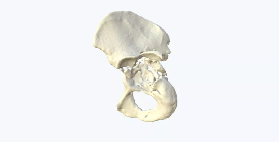
Case A patient presented with severe trauma following a motorcycle accident which resulted in an associated both column fracture of the acetabulum. Due to the severity and comminution of the fracture, 2D imaging did not convey the full extent of the fracture pattern and fragment location. This made it difficult to conceptualize the pathology. The […]
Confirming the right course of treatment and improving the patient consent process with physical 3D models

The Case Dr. Michael Pearl is an Orthopedic Surgeon with a specialty in shoulder and elbow surgery. When researching for a case regarding a bone deformity, he found that the literature he consulted was opposing his own instincts about the treatment. To gain more clarity, he had a 1:1 anatomical 3D model created, specific to […]
3D Printed Model Enhances Pre-Operative Planning for Complex Calcaneal Fracture
A male patient was admitted to Ulster Hospital with severe injuries to his left calcaneus due to a crushing incident. The intricate and closely positioned anatomical structures within the foot made it challenging to obtain a comprehensive assessment of the extent of the injury using conventional CT scans. Solution The surgical team turned to Axial3D […]
Transforming visualization for scoliosis surgery planning

All back surgery is stressful and worrying for patients. However, these concerns are often multiplied with scoliosis surgery, that brings additional layers of complexity to the operating table. The surgery typically requires the clinical team to painstakingly align curved vertebrae so that they heal into a single, solid bone – and with utmost precision, otherwise […]
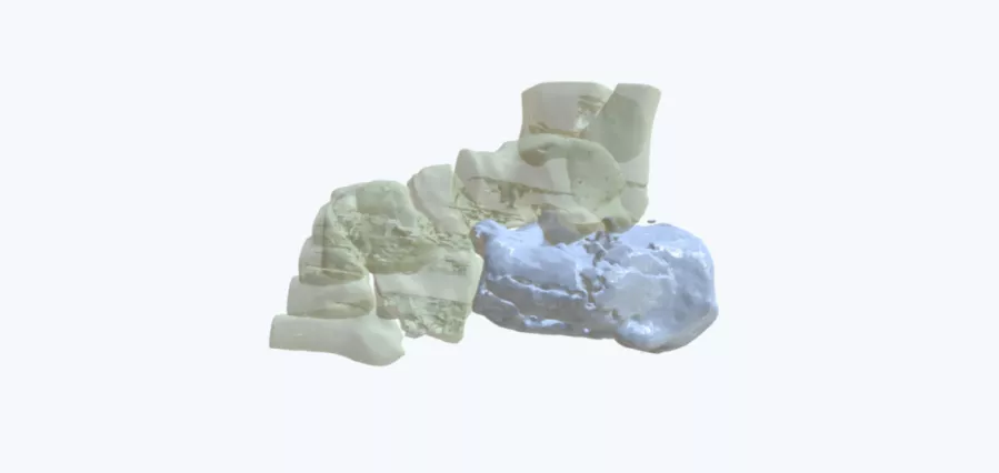
When 2D imaging doesn’t cut it: 3D modeling for enhanced insights
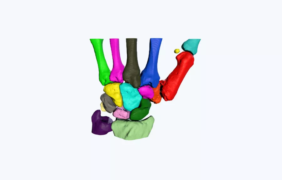
Case A 3D printed model was used to improve the understanding of the size and morphology of a deformed trapezium. Due to the complex anatomical arrangement of the carpal bones with the articulating aspects of the metacarpals, the understating of the anatomy of the bone was extremely difficult to conceptualize on CT scans alone. The […]
