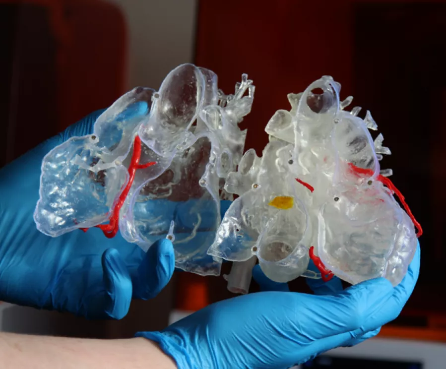
Improving patient safety in surgery with a 3D printed cardiac model

No surgery comes without risk, but some procedures will naturally carry an increased level of risk than others. Take cardiac surgery as an example, where the complexity of many cases can increase the potential for causing significant harm to the patient. Thankfully, we have seen incredible advances in cardiac surgery knowledge, techniques, equipment, and skill […]
Using patient-specific models to plan complex congenital deformity corrections (PAPVR)

Dr. Jenny Zablah is the Assistant Professor of Pediatrics Interventional Cardiology who uses Axial3D models within the specialized cardiac catheterization laboratory, where they treat children and adults, with congenital heart disease. In this case, the patient in his twenties has complexities causing the pulmonary veins to not connect to the left atrium and connect instead […]
3D anatomical model saves time and reduces risk on a rare transcatheter pulmonary valve replacement
Case In preparation for a percutaneous pulmonary valve implantation, or transcatheter pulmonary valve replacement, a 44-year-old woman required a procedure involving the replacement of the pulmonary valve through catheterization via a vein. Solution The complexity of the planned procedure prompted the surgical team to opt for a 3D anatomical model. This advanced tool provided the […]
Supporting faster and safer cardiac valve replacement with 3D modeling

Approximately eight out of every 1,000 babies are born with congenital heart disease (CHD). Typically, babies born with these complex cardiac anomalies will undergo surgical treatment in the first weeks or months of life, but even with this, many will require further procedures well into adulthood. The Case In this case, surgeons treating a 32-year-old male with […]
Helping a cardiac surgeon to visualize their own ASD with 3D printing

As a child, Dr. Furdova was diagnosed with septal defect following some check-ups on her heart. Small atrial septal defects can spontaneously close during childhood, however this is not always the case. “During my studies I became very interested in cardiac surgery. Before taking on a new position, during a routine check-up, my GP referred me to […]
3D printed model used to assist in the planning of a transcatheter balloon septostomy
Case A male patient who had traveled to the UK from Syria presented with a complex form of congenital heart disease – double inlet left ventricle, with transposed great vessels and pulmonary atresia. There was a degree of left atrio-ventricular valve atresia. He was status post initially aorto-pulmonary shunt, with subsequent take down and creation of […]
Enabling a 60-minute reduction in pediatric surgery with a 3D printed anatomical model
The Case A baby girl presented with atrioventricular (AV) and ventriculoarterial (VA) discordance – a complex type of heart lesion that accounts for less than 1% of infants with congenital heart disease – alongside a ventricular septal defect (VSD) with pulmonary stenosis (PS). 2D imaging made it difficult for her surgeon to visualize the full […]
Aortic valve surgery made safer and faster
The Case In this case, a 58-year-old man suffering from aortic stenosis and a recessive right coronary artery (RCA) needed an aortic valve replacement. Aortic stenosis, a narrowing of the aortic valve opening, is one of the most common and serious valve disease problems which restricts the blood flow from the left ventricle to the […]
3D Printed Left Atrial Appendage Model Aids Pre-Operative Planning and Patient Consent
Case A patient presented with a complex condition involving their Left Atrial Appendage (LAA). The precise classification of the LAA’s abnormality was proving to be challenging based solely on the information obtained from CT scans. Recognizing the need for a clearer understanding of the problem, the surgeon requested a 3D printed anatomical model. Outcome Turning […]
