Revolutionizing Pediatric Cardiac Care at Southampton Hospital

In the heart of Southampton, United Kingdom, Mr. Nicola Viola, clinical lead for congenital cardiac surgery, and Dr. Tara Bharucha, foetal and pediatric cardiologist, have dedicated years to introducing 3D technology to help improve surgical planning, firmly believing it’s an indispensable tool for any surgeon who experiences its capabilities firsthand. By incorporating 3D printed models […]
Enhancing Surgical Precision: Patient-Specific 3D Model for Vascular Calcification Assessment
In vascular surgery, the precision of clamping for complex surgeries such as organ transplantation is pivotal to safeguard the vessel and ensure the procedure’s ultimate success.Clamping in areas with pronounced calcification may lead to insufficient vessel occlusion post-clamping, compromising the procedure’s efficacy. The significant challenges posed by vascular calcification in major vessels necessitate meticulous pre-operative […]
Optimizing surgical outcomes with 3D models in Double Outlet Right Ventricle (DORV) cases

Dr. Ala Al-Lawati, Head of the Cardiac Surgery Department at the National Heart Centre of the Royal Hospital in the Sultanate of Oman, uses Axial3D solutions in the field of paediatric cardiac surgery. Case A patient diagnosed with Double Outlet Right Ventricle (DORV) and multiple Ventricular Septal Defects (VSDs) presented a challenging scenario for surgical […]
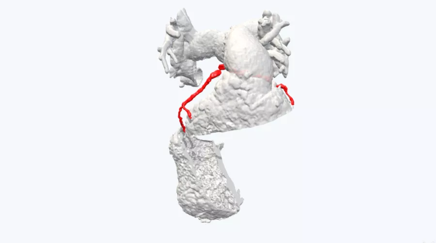
Gaining insights into anatomical intricacies of ccTGA using patient-specific 3D solutions

Dr. Ala Al-Lawati, Head of the Cardiac Surgery Department at the National Heart Centre of the Royal Hospital in the Sultanate of Oman, uses Axial3D solutions in the field of paediatric cardiac surgery. Case This case study presents a patient with Congenitally Corrected Transposition of the Great Arteries (ccTGA), Ventricular Septal Defect (VSD) and […]
Getting to the safest surgery plan

Around 1 in every 240 people are born with a ventricular septal defect (VSD) each year in the United States. For most people, the causes of heart defects such as a VSD are unknown and over time, if not repaired, this defect can increase the risk for other complications, including heart failure, pulmonary hypertension, or […]
Medical 3D model saves crucial time in heart transplant surgery for a patient with congenital heart disease

Abdel Hakim Moustafa is a Cardiologist and specializes in advanced cardiac imaging. He works at Hospital Sant Pau in Barcelona where they mainly focus on cardiovascular diseases. In the video below, he talks about a recent case in which a patient with congenital heart disease needed a heart transplant. Every day, Dr. Moustafa works with […]
Clinical team uses 3D printed model to reduce risk in cardiac surgery

Congenital heart defects are the most common human birth defect, found in up to 2% of the population. Case A patient presented with post-repair of pulmonary atresia with ventricular septal defect requiring pulmonary valve replacement; due to a stenosed RV-PA conduit. Given their history of multiple previous cardiac surgeries, a further surgery would be of […]
3D printed anatomical model transforms surgical plan and informed consent process in pediatric RVOT case
A 15-year-old female patient presented with a condition known as right ventricular outflow tract (RVOT) tachycardia. RVOT ventricular tachycardias (VT) manifest without underlying structural heart issues and are classified as idiopathic ventricular arrhythmias. These arrhythmias are believed to arise from adenosine-sensitive, cyclic AMP mediated, triggered activity, and are prevalent among adolescents and young adults. (Source: Abdel […]
3D cardiac model supports Hancock valve replacement

Case A 20-year-old female presented with pulmonary atresia with VSD. A previous Hancock conduit required revision and stenting. It was difficult to assess the distance of the replacement site with coronary branches, so the surgeon requested a 3D printed model to augment the 2D scans. The CT scans were used to create a 1:1 scale […]
Correcting Transposition of the Great Arteries with the Help of a Patient-Specific 3D Printed Anatomical Model
Case Dr. Nicola Viola is a Cardiac Pediatric Surgeon at University Hospital Southampton NHS Foundation Trust. In one of his recent transposition of the great arteries cases, surgical planning proved challenging due to the size of the patient’s heart. The patient presented with transposition of the great arteries with stenosis of the pulmonary artery, […]
