Case
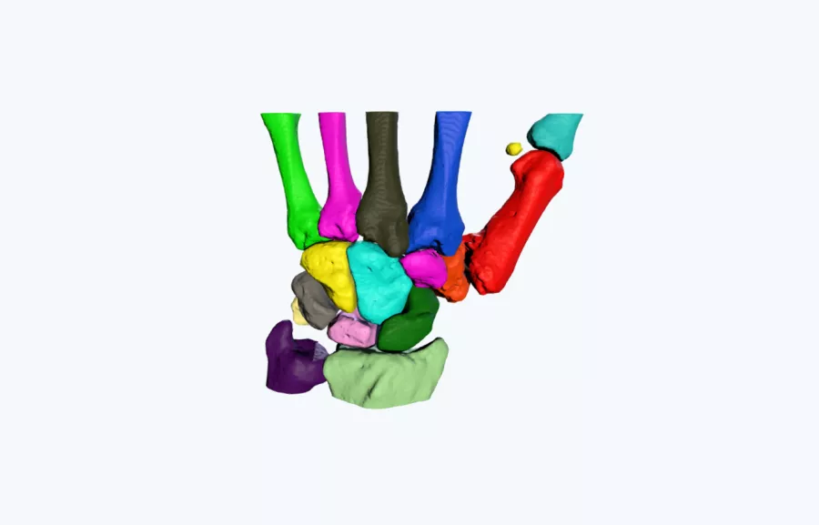
A 3D printed model was used to improve the understanding of the size and morphology of a deformed trapezium. Due to the complex anatomical arrangement of the carpal bones with the articulating aspects of the metacarpals, the understating of the anatomy of the bone was extremely difficult to conceptualize on CT scans alone.
The 3D printed anatomical model enabled the surgeon to gain a much greater insight into the shape of the bone, which improved his confidence that surgery was indeed the correct approach.
Outcome
“Having the 3D printed model allowed me to see the whole deformity on the trapezium which was not possible with the original CT scans provided. The pathology was much more complex than initially diagnosed, confirming my decision-making that surgical intervention was required.”
– Mr Jeremy Field, Consultant Orthopaedic and Hand Surgeon, Cheltenham General Hospital, Gloucester
