Case
Shoulder anatomy presents an intricate challenge for surgeons, characterized by a complex network of overlapping and articulating structures. Navigating surgical planning for these intricate 3D structures proves exceptionally challenging when relying solely on 2D images.
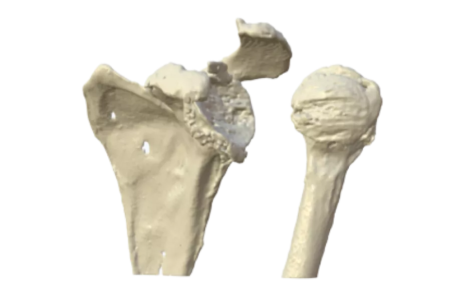
In this case, the patient exhibited a complete fusion of the humeral head and glenoid fossa, resulting in a complete loss of joint mobility.
Outcome
Surgeon, Mr. Eames, proactively engaged Axial3D to generate multiple 3D printed models derived from the patient’s 2D CT scans. This approach facilitated a comprehensive assessment of bone stock in both the humeral head and glenoid fossa. Furthermore, it empowered Mr. Eames to evaluate the joint structure post-separation.
The 3D models emerged as indispensable tools in Mr. Eames’ meticulous plan for separation and reconstructive procedures. This innovative technology allowed him to precisely strategize his surgical approach and gather the requisite equipment for reconstruction well in advance of entering the operating theater. The result was a streamlined surgical process, reducing time spent in the operating room and significantly enhancing the overall patient experience. Subsequent to the procedure and joint replacement, the patient regained use of their shoulder joint, marking a substantial improvement in their quality of life.
Mr. Eames emphasized the pivotal role of the 3D printed model, highlighting its ability to provide an unparalleled understanding of the intricate arrangement of the glenohumeral joint. He acknowledged that such a level of comprehension would have been unattainable with the conventional imaging tools currently at his disposal.
“The 3D printed model gave a much greater understanding of the complex arrangement of the glenohumeral joint. I just wouldn’t have been able to get this level of understanding from my current imaging available to me.”– Mr. Michael Eames, Consultant Orthopedic Surgeon, Ulster Hospital, Belfast
