Case Study18th January 2024
Revolutionizing Pediatric Cardiac Care at Southampton Hospital
Transforming reconstructive surgery following removal of jaw bone tumor
Case Study
Squamous cell carcinoma (SCC) is the most common form of malignancy in the head and neck. Quite often an SCC will imitate an infectious condition when viewed on 2D radiography, posing a diagnostic challenge for even the most experienced clinicians.
The Case
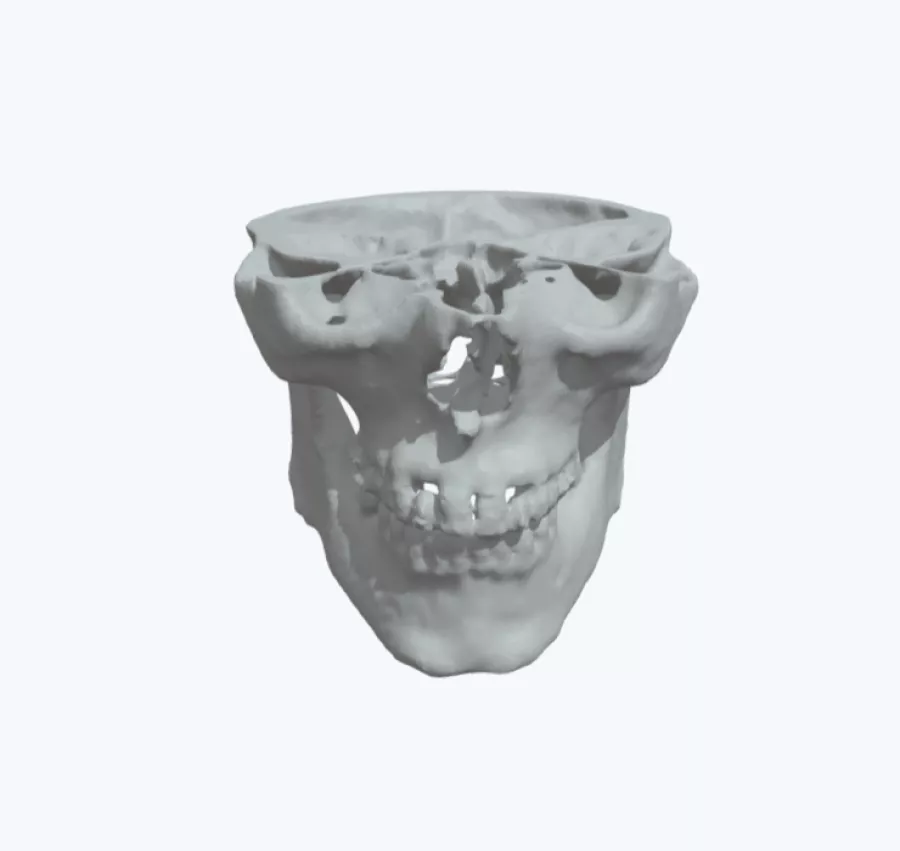
In this case, doctors treating a 64-year-old man found what they believed could be an SCC in the right side of his jaw but couldn’t be sure, meaning they would have to plan for any and all possibilities they may find once they started operating. To remove this uncertainty in advance, the lead surgeon ordered a 3D model of the man’s skull and jaw from Axial3D.
Solution
With the 3D model at their disposal, the clinicians were able to confirm that he was indeed suffering from an SCC in his jaw and set about with full certainty defining their surgical plan to remove the tumor, along with a portion of his jaw bone. The team then planned to rebuild his jaw with reconstructive plates.
Benefits of using the 3D Model
Having fulfilled its original purpose in enabling the surgeon to clarify the patient’s condition and define a safe surgery plan, the team then found an incredible secondary use for the model. Using it as a fitting guide, they pre-bent the reconstruction plates ahead of time, resulting in an impressive reduction of sixty minutes of time spent in the OR. There is growing evidence to show that using 3D models to pre-bend plates can not only save time in the OR, as it has done in this case, but also offer better fitting accuracy too. This has a huge impact on the patient’s recovery and rehabilitation, including recovering their self-esteem, and significantly reduces their risks of downstream complications.
Have a case you would like to discuss? Talk to an expert to learn how a patient-specific model can help you improve outcomes.
Contact us
Case Study8th November 2023
Enhancing Surgical Precision: Patient-Specific 3D Model for Vascular Calcification Assessment
Case Study28th August 2023
Optimizing surgical outcomes with 3D models in Double Outlet Right Ventricle (DORV) cases
Case Study28th August 2023
Gaining insights into anatomical intricacies of ccTGA using patient-specific 3D solutions
Case Study2nd December 2021
Correcting Transposition of the Great Arteries with the Help of a Patient-Specific 3D Printed Anatomical Model
Case Study29th November 2021
Medical 3D model saves crucial time in heart transplant surgery for a patient with congenital heart disease
Follow us


