The Case
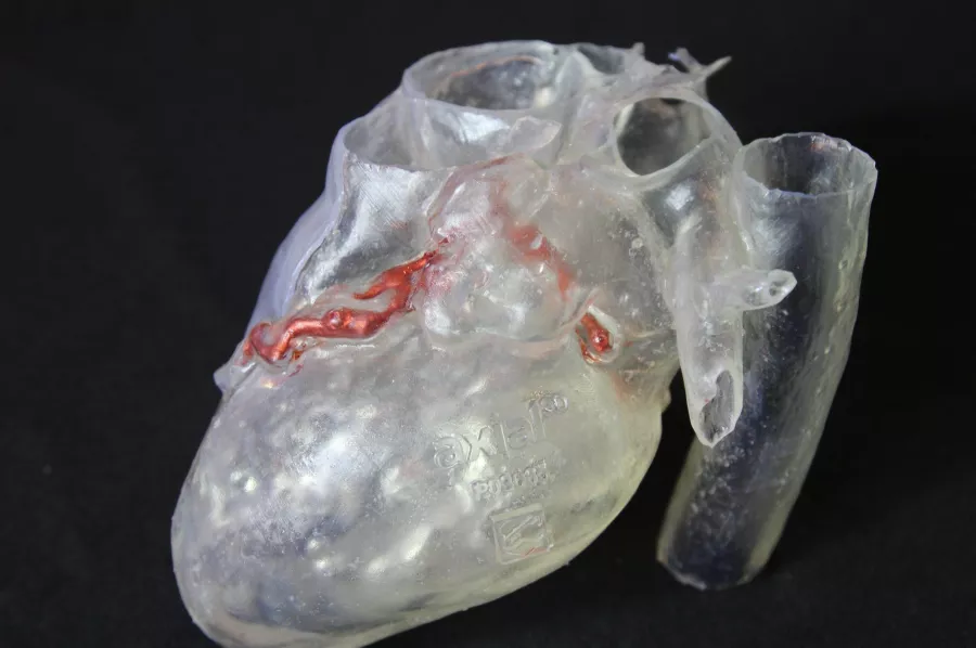
In this case, a 58-year-old man suffering from aortic stenosis and a recessive right coronary artery (RCA) needed an aortic valve replacement. Aortic stenosis, a narrowing of the aortic valve opening, is one of the most common and serious valve disease problems which restricts the blood flow from the left ventricle to the aorta.
Solution
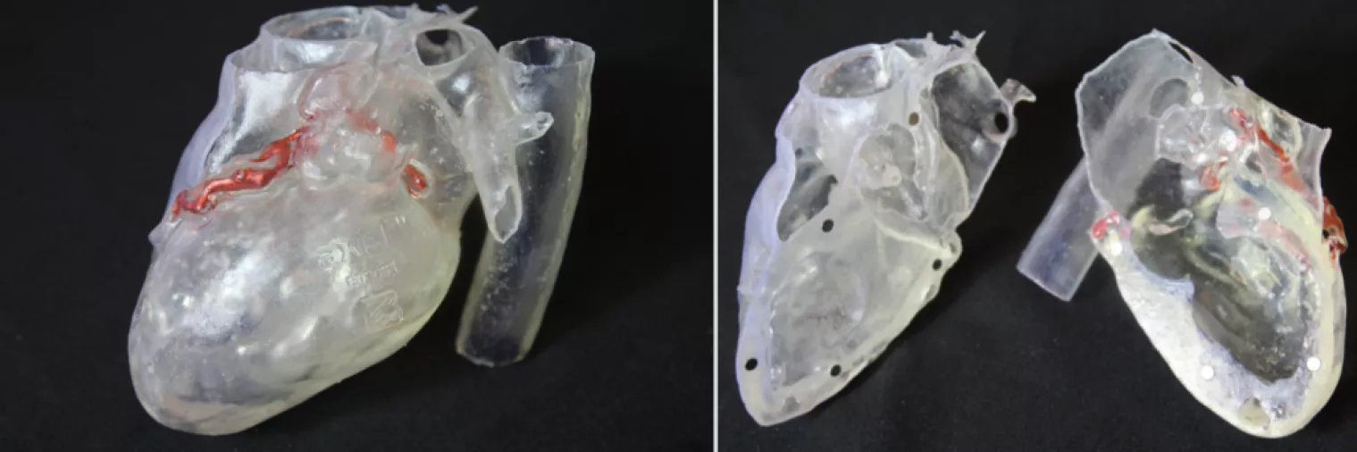
The lead physician requested a clear model of the patient’s myocardium with oversized coronary arteries to allow him to plan his procedure by injecting resin into the coronary artery to better understand how the patient’s heart was being impacted. He also hoped to allow inspection of the valve above and below and visualization of the coronary arteries using the model.
Disclaimer: Details of Axial3D’s regulatory clearance for diagnostic use cases are outlined here. For all other uses of Axial3D solutions, they should be used for demonstration and education purposes only.
